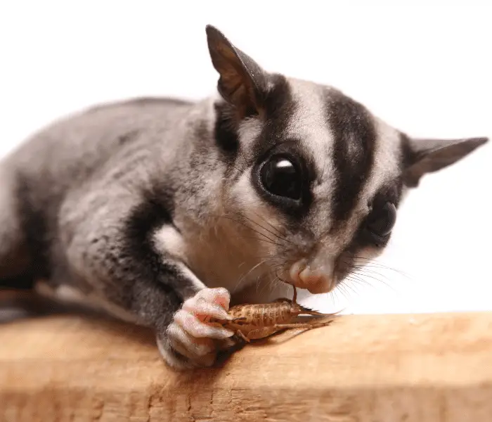
Sugar gliders are nearly as good at stealing hearts as they are at seeing in the dark. We are not entirely sure whether sugar gliders see colors the same way we do, but we know that they have an acutely keen vision to spot insects and fruits from a distance.
Sugar gliders also have a vast field of vision which helps them to be able to spot predators in the wild.
Eye conditions are one of the most common health conditions sugar gliders suffer from (second to obesity). This article aims to inform sugar glider owners on a few of the most common eye conditions of sugar gliders and possible causes and treatments for these conditions.
Eye Structures
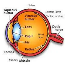
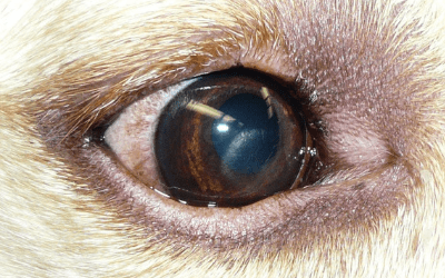
The picture of the eye below serves as a reference image to better understand the eye conditions discussed below. Important structures to take note of are the cornea (corneal ulcers and corneal lipidosis), the lens (cataracts), and the tapetum lucidum (a reflective part at the back of the eye that helps animals see at night – sometimes confused with cataracts and corneal lipidosis).
Corneal Ulcers
Corneal ulcers are erosions or small breaks in the eye’s outer layer (the cornea), commonly caused by a scratch to the eye, often inflicted by sharp objects in the cage or by cage mates.
Sugar gliders are prone to getting corneal ulcers due to their shallow eye sockets and protruding eyes. You might not be able to see the scratch on the eye, and often the only early symptoms of an ulcer are runny eyes and squinting.
Deeper ulcers often become infected if left untreated, which causes the erosion to become wider and deeper, and the infection to spread to the soft tissue surrounding the eye. A deep or infected ulcer is very painful and needs urgent veterinary care. Your vet can diagnose a corneal ulcer by applying a stain to the eye.
If the ulcer has become infected, you might notice a white or yellow discoloration on the eye’s outer layer, often surrounded by a blueish discoloration of the surrounding cornea (corneal edema).
Blue discoloration of the cornea (outer layer of the eye) is due to swelling or accumulation of fluid in that area, called corneal edema. Corneal edema is usually a symptom of certain eye conditions and the underlying cause should be identified.
Corneal ulcers are commonly treated using topical antibiotic eye drops and pain medication. However, if urgent and aggressive treatment has not ensued, the corneal ulcer may progress to form an eye abscess.
At this point, the affected sugar gliders are often systemically ill, and the eye can no longer be saved, in which case surgery to remove the eye may be the only remaining option.
Care should be taken to prevent future corneal ulcers. For example, remove cage accessories with sharp or protruding points or separate gliders who do not get along.
Cataracts
Cataracts are a common eye condition of sugar gliders and may be identified as a white spot in the lens of the eye (check the eye anatomy diagram above to see the lens).
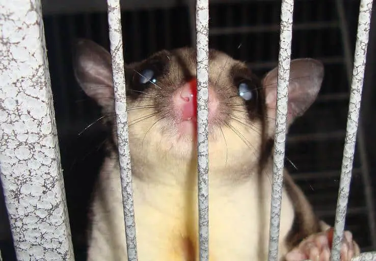
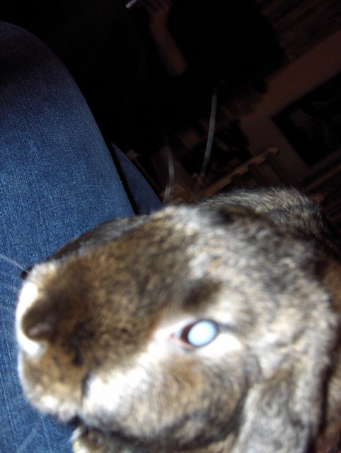
Cataracts are sometimes confused with the tapetum lucidum, a normal structure in animal eyes that helps them to be able to see at night. When light is reflected off the tapetum lucidum, it appears white, similar to a cataract. In addition, corneal lipidosis (discussed below) may also sometimes look like cataracts at first glance.
Cataracts are sometimes confused with the tapetum lucidum, a normal eye structure and with Corneal Lipidosis, which is an abnormal accumulation of fatty crystals in the outer layer of the eye. All three of these have a greyish appearance but have very different causes.
Cataracts cause partial vision loss in most cases but may cause complete loss of vision in severe cases. Depending on the cause of the cataract, vision loss may be reversible.
Depending on the severity and cause, surgical removal of cataracts may be possible by veterinary ophthalmologists; however, this is a high-risk surgery, and glaucoma is a common complication after this surgery in sugar gliders.
Causes of Cataracts in Sugar Gliders
1. High Carbohydrate Diet
The lens of the eye is very sensitive to the amount of sugar in the blood (blood glucose). This is, for example, why diabetic humans are more prone to developing cataracts. A similar mechanism is seen in sugar gliders on a high-sugar, high carbohydrate diet, as most marsupials do not cope well with hyperglycemia (Johnson-Delaney, 2002).
Cataracts seem to be more common in adult gliders who eat a diet high in corn, fruits, and sugar such as honey. To learn more about the ideal diet for sugar gliders (the way wild sugar gliders eat), you can have a look at this article.
There is no evidence to suggest that cataracts in sugar gliders are caused by a high fat diet.
2. Hand Reared Sugar Gliders Fed Cow’s Milk
Sugar gliders, and all marsupials for that matter, are unable to completely metabolize the sugar in cow’s milk (lactose). This causes the accumulation of a metabolite called dulcinol in the lens of the eye, which pulls fluid into the lens, causing it to become opaque.
If cataracts in joeys are picked up early, and a diet change to a milk substitute that does not contain galactose is made, these cataracts may be reversed; however, this may not always be the case.
3. Congenital
Congenital conditions are conditions seen in newborn or young animals and are usually due to genetic or developmental abnormalities.
Congenital cataracts are often seen in joeys from a dam that has been pushed for breeding or has been on an inadequate diet, often deficient in protein and high in sugar.
Breeding females will often have yeast or fungal pouch infections, and it was proposed that this could contribute to cataracts in joeys as well.
If other gliders in the lineage have a history of cataracts. A genetic component is a likely cause, and that particular lineage should not be used for further breeding.
Corneal Lipid Infiltrations (Corneal Lipidosis)
Corneal lipidosis is white fatty accumulations in the eye’s outer layer (the cornea), often seen in youngsters from does who are fed a diet excessively high in fat. Corneal lipidosis may also be seen in adults who eat a high-fat diet.
Corneal lipidosis appears as white, sometimes sparkly, or crystalline material in the cornea and will typically resolve when appropriate diet changes are made.
Conjunctivitis
Conjunctivitis is the term used to describe infection and inflammation of the soft tissue surrounding the eye and the transparent membrane covering the white part of the eye.
Conjunctivitis can be primary when caused by a bacterial or viral infection or secondary when it is a result of another eye condition such as glaucoma.
If primary, conjunctivitis is treated using anti-inflammatory pain medication and antibiotics (eye drops or oral). When conjunctivitis is secondary to another eye condition, conjunctivitis will typically resolve when the inciting cause is addressed; for example, the glaucoma is treated.
Glaucoma
Glaucoma is an eye condition where the pressure inside the eyeball is abnormally high. This can be due to many different causes of which some are treatable and others not.
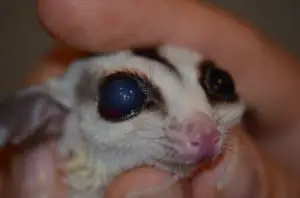
Glaucoma is diagnosed by measuring the pressure inside the eye (the intraocular pressure or IOP) with a special device that bounces against the eyeball. Not all vets have access to this device, though. In advanced cases of glaucoma, the eyeball will appear enlarged. This may be easier to notice if only one eye is affected.
The eye continually produces ‘eye fluid’ that circulates inside the eyeball. As new fluid is produced, old fluid needs to drain. Glaucoma develops when there is a problem with the overproduction of the fluid or when there is a problem with the draining of the fluid. For example, if there is bleeding inside the eyeball (let’s say due to the eye taking a hard knock), small blood clots can block the draining area, causing over-accumulation of fluid in the eye.
Early symptoms of glaucoma include squinting and whitish discoloration of the eye (corneal edema). Glaucoma is very painful, and a high IOP for an extended period of time will cause permanent blindness.
Retrobulbar Abscess
A retrobulbar abscess is a pocket of infection behind the eyeball. In sugar gliders, this is commonly due to a gum and tooth infection ascending up the tooth root until it infiltrates the pocket behind the eye.
This condition is extremely painful and is characterized by swelling around the eye and one eye slightly protruding (this condition rarely occurs in both eyes at the same time).
Again, diet plays a large part in preventing these conditions. Gliders fed a diet high in sugar are prone to dental disease and tooth decay. Feeding a diet that includes insects with an exoskeleton(crunchy outsides) such as mealworms can help keep teeth clean.
Taking your sugar glider for an annual check-up to pick up on tooth problems early will be helpful. A proper mouth exam is best done while your glider is sedated. Your vet will also be able to clean the teeth and extract any possible rotten teeth to prevent abscesses from forming.
Signs Of A Painful Eye
1. Squinting
If your glider is keeping their eye shut or half-shut or seems unable to open their eye fully, it is an indication that the eye is uncomfortable or sensitive.
2. Runny Eyes
Irritated or painful eyes will produce excess clear or watery tears.
If you see a white, yellow, or greenish discharge, an infection is present, and an urgent vet visit is indicated.
3. Rubbing The Eye
Sugar gliders may rub or paw at their eyes if it is painful. If they constantly paw at their eyes, they may cause further damage to an already sore eye.
Eye Conditions Need Urgent Veterinary Attention
If your sugar glider shows symptoms of eye discomfort lasting longer than one day, you should consult your vet urgently. Eye conditions are always time-sensitive as your sugar glide’s vision may be at stake.
Sugar gliders are also very good at hiding symptoms, so by the time your glider seems visibly ill, they are often life-threateningly sick!
Should I Let My Sugar Glider’s Eye Be Removed?
Unfortunately, there may be some situations where removing a diseased eye will be the best option for your glider. Examples of such cases include:
- The eye is not responding to treatment and is severely painful
- Permanent vision loss has already occurred, and there is an infection present
- The risk of complications associated with eye-salvaging surgery outweighs the risk of eye removal surgery
- Specialist treatment can not be pursued due to many reasons such as accessibility or costs, and the eye is causing discomfort to the sugar glider
- There is cancer present in the eye and removal will limit the spread of cancer.
Most animals can adapt surprisingly quickly after the removal of an eye. Their other instincts kick in to help them navigate their environment. They are not worried about the aesthetics of only having one eye. As long as they can be pain-free, comfortable, and cared for, they will be happy.
References
- Banks, R., 2013. Exotic small mammal care and husbandry. Hoboken: John Wiley, pp. 157 – 168. https://onlinelibrary.wiley.com/doi/book/10.1002/9781119265405
- Dierenfeld, E., 2009. Feeding Behavior and Nutrition of the Sugar Glider (Petaurus breviceps). Veterinary Clinics of North America: Exotic Animal Practice, 12(2), pp.209-215.
- Fraess, GA, Sadar, MJ, Daniels, JB, Sharkey, L, Henriksen, MDL. Clinical ophthalmological diagnostic description of 10 healthy sugar gliders (Petaurus breviceps) and prevalence of ocular-related presentations in a larger hospital population. Vet Ophthalmol. 2021; 24: 80– 92. https://doi.org/10.1111/vop.12850
- Meredith, A. and Redrobe, S., 2002. BSAVA manual of exotic pets. 4th ed. Quedgeley: British Small Animal Veterinary Association, pp. 102 – 106 25. https://www.researchgate.net/publication/240493099_The_BSAVA_Manual_of_Exotic_Pets_4th_edn
